Overview
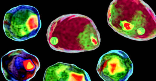
The study of biological structures from cells to tissue are critical to advancing the sciences of pharmacology, biology, epidemiology, immunology, pathology, and many more. Whether the target is unicellular or studying intracellular events or thick tissue, having a precision light source for medical imaging is critical to the success of even the most advanced optical arrangements. Modern studies rely on capturing as much information as possible while maintaining the integrity of the specimen being examined. Having exact control over the light source allows for intensity control while holding color temperature with high fidelity and very high intensity to reduce the exposure time and limit phototoxicity. Light matters.
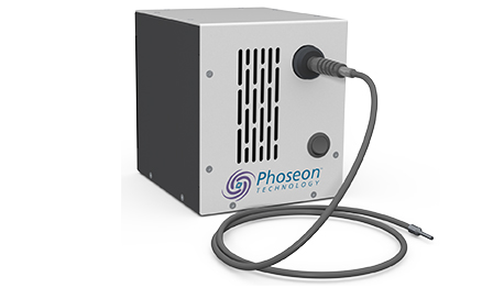
KeyLight™ illumination systems from Phoseon offer OEMs reliable, high-performance imaging for laboratory instruments, automated microscopy instruments, medical device illumination, and high content screening applications. KeyLight delivers intense, broad-spectrum UV and visible wavelengths for a wide variety of colors between 340nm and 760nm. These compact light sources support 3, 4, 5 or 6 channel systems with customizable wavelengths and UV intensity for greater flexibility depending on the application requirements, and are easily integrated into OEM systems. The product form factor and control options are designed specifically to support each type of OEM instrument.

KeyLight Benefits
- Small form factor for easy integration
- High performance imaging
- Cost and energy savings
- Compact architecture supports various optical fiber sizes
- Optimized for various numerical aperture requirements
- Low maintenance with mercury-free operation
- Precision output control
- Color temperature selectability
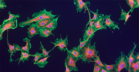
Medical Imaging Applications
- Live cell imaging
- Cell culture
- Thick tissue
- Tissue autofluorescence
- Fluorescence
Other Application Areas
- Oil immersion microscopy
- Kohler illumination
- Darkfield microscopy
- Differential interference microscopy
- Phase contrast microscopy
- Zebrafish research